
Steps to Lower Your Risk for Breast Cancer
- Get regular screenings
- Maintain a healthy weight
- Be physically active
- Limit alcohol consumption
- Avoid smoking
- Manage stress
- Know your family history
For more information, visit Five Ways to Help Reduce Your Breast Cancer Risk | American Cancer Society

Screening Mammogram
ACR recommends annual mammography beginning at age 40.
STRIC offers DBT (Digital Breast Tomosynthesis) commonly referred to as 3D for both screening and diagnostic exams.
What are the advantages of having DBT with C-view?
DBT imaging takes picture of the breast in very thin slices which eliminates tissue overlap. These thing slices allow the radiologist to view a clearer image of the inner breast tissue, an important benefit for dense breast tissue.
C-view is an advanced technology that creates an image that is beneficial for comparison to your prior mammogram. The C-view image adds no radiation exposure to the low dose DBT exam.
What to wear?
Two piece outfit and comfortable shoes are preferred. You will undress from the waist up and change into a gown. Do not wear perfume, powders or deodorant as it can cause artifacts on the images.
What happens during a screening mammogram?
A screening mammogram consists of four (4) views, two views of each breast.
The technologist will reposition you for each view and apply compression to flatten and even out the breast tissue for better visualization and imaging.
After the images are taken, the technologist will review them for positioning and clarity. If there is any movement or breast tissue missing from image, she will repeat the image.
Once the images have been reviewed by the technologist, they are sent electronically to our PACS system to be read by one of our board certified radiologists.
What if I have breast implants?
Mammography for implants is considered a routine annual screening procedure. If you have any breast symptoms or changes you will need to contact your physician to obtain an order for a Diagnostic Mammogram.
What if I notice a change in my breast(s) when I perform my monthly breast self-exam?
If you feel or see something unusual such as thickening, nipple discharge, pain or a lump call your doctor immediately, and he/she will determine your next course of care. If you have any of the above, you do not qualify for an annual, routine screening mammogram. Please contact your physician to obtain an order for your Diagnostic Mammogram.
Do I qualify for a Screening Mammogram if I have a prior history of breast cancer?
If you have a prior history of breast cancer, but have remained stable with no new symptoms, you may be considered for a Screening Mammogramm. Please contact your physician prior to scheduling your appointment to see if you qualify.
What happens next?
Your results will be sent to you and your healthcare provider. If your exam is normal, you will need to do nothing further and we will see you next year! We will remind you when it is time for your annual mammogram the following year. You should keep up with your breast self-exams monthly and report any changes to your healthcare provider.
If your screening shows an area that cannot be determined to be normal or stable, you will be asked to return for additional imaging at a STRIC location that specializes in Diagnostic Mammography. In most cases, the work-up exam will be a diagnostic mammogram with views to magnify the area/and or give more localized compression for better detail. Ultrasound is commonly uses to gain more information. If either or both of these are recommended, we will inform you in your results letter and included a pre-scheduled appointment for you to return. If the day or time is not convenient you mat contact us to reschedule.
What will my experience be like if I am referred for further exams or work up?
You will be referred to a STRIC location specializing in Diagnostic Mammography. Radiologists and staff are specially trained in breast imaging to provide you with the highest level of care possible. Because of the level of care and service, the necessity of tailoring each exam to your individual problem, and the involvement of the radiologists, you may experience a wait time associated with your exams.
Mammography AI
Your mammogram is interpreted by board-certified, radiologists who use Artificial Intelligence as a tool to assist in the reading. AI technology detects suspicious areas that may not be visible to the human eye and marks them for the radiologist to review. Artificial Intelligence, combined with highly trained radiologists allows for earlier detection of breast cancer. To learn more about how AI is revolutionizing mammography and early breast cancer detection, check out: AI is the Next Step Forward in Early Breast Cancer Detection.
Breast Density
The results of your mammogram will include information on your breast density classification. Breast tissue can be either dense or not dense. Dense tissue makes it harder to find breast cancer on a mammogram and also raises the risk of developing breast cancer. You will be informed if your breast tissue is considered dense or not dense. If you have dense breasts, you should speak with your healthcare provider about your individual situation and the options available to you for additional imaging. For more information on breast density click here.
Diagnostic Mammogram
What is the difference between screening and diagnostic mammogram?
Screening mammography is intended for women 40 and over that have no symptoms. Diagnostic mammography is for women who have symptoms such a pain, lump, discharge or dimpling or who have been asked to return for additional imaging as a result of their screening mammogram.
What happens during the exam?
A radiologist that specializes in mammography will inform the technologist which specific views to take to best visualize the area. These views are taken on the same equipment as the screening mammogram. Compression will again be used as well as specialized views and/or magnification of the area where the radiologist needs to see more detail
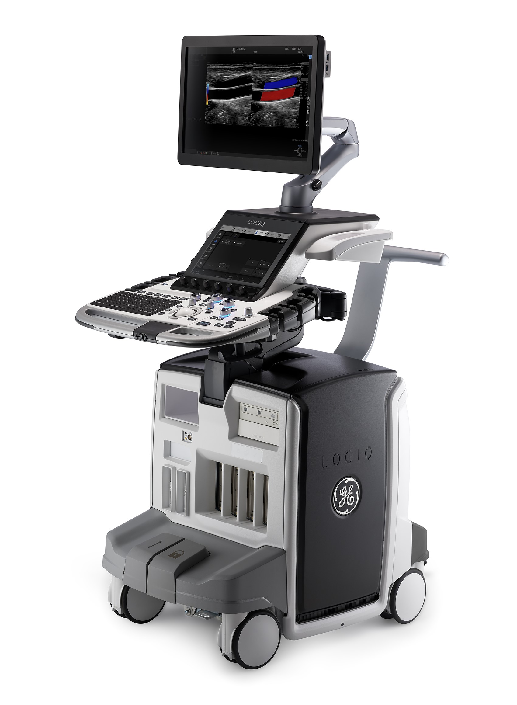
Breast Ultrasound
What is ultrasound used for?
Breast ultrasound is used to help the radiologist diagnose a breast lump found on a self or clinical breast exam or a nodule identified on a mammogram. This imaging will show if the finding is fluid-filled (such as a benign cyst), a solid mass (which may need further testing to ensure it is not abnormal) or a lump that is both cystic and solid. It can identify other abnormalities in the breast or axilla seen on the mammogram. It is also clinically indicated as an additional tool for patients that have dense breast tissue (tissue that has more glandular & connective tissue and not very much fatty tissue).
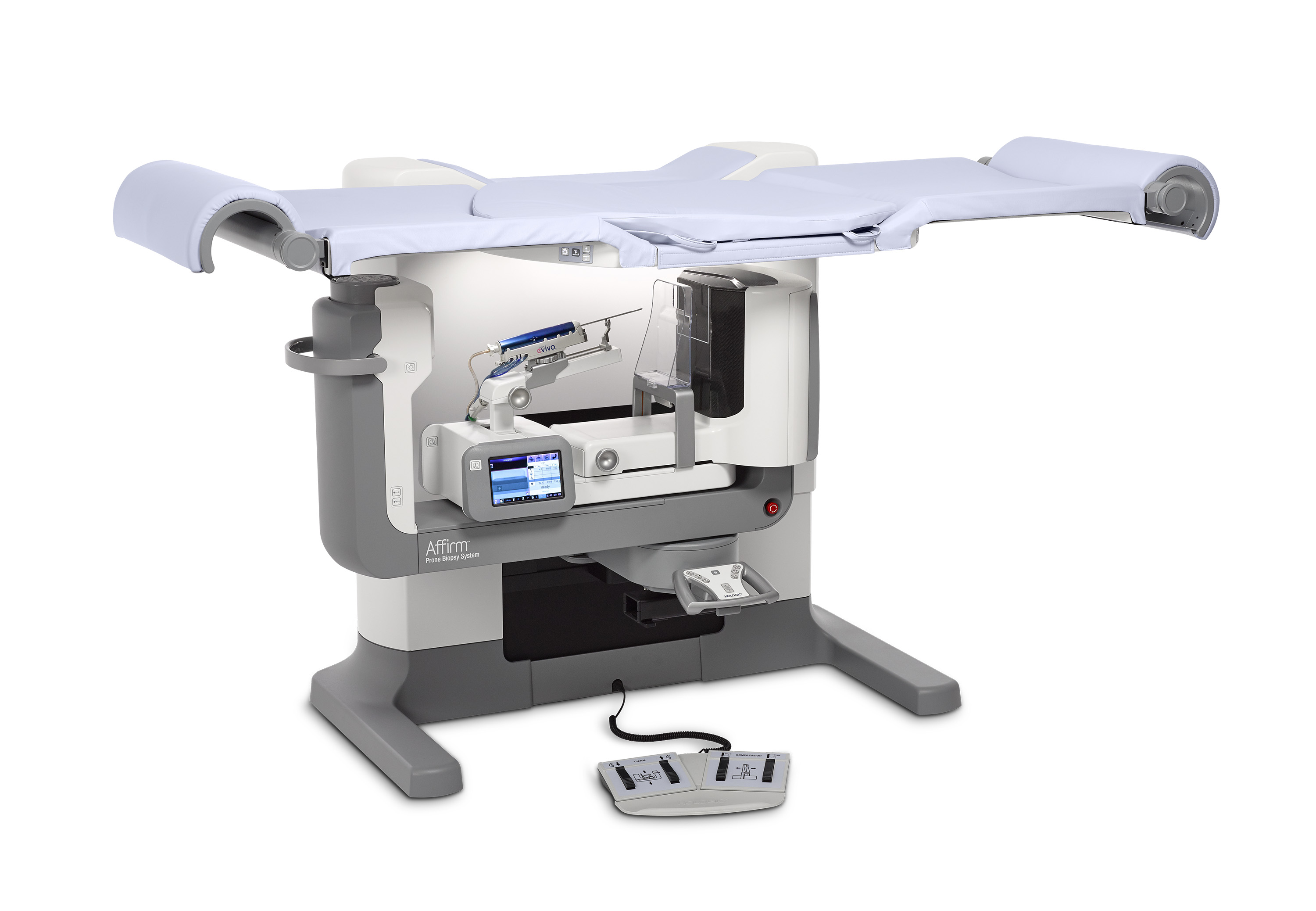
Breast Core Biopsy
What is a core biopsy?
A minimally invasive procedure that uses imaging equipment, either stereotactic or ultrasound, to locate the area to be evaluated. The radiologist uses the equipment to guide the biopsy needle to the area and takes a tissue sample to send to the laboratory.
What should I expect during procedure?
You will first speak with the radiologist about what to expect during the procedure and to answer any questions you may have. For a stereotactic biopsy you will be positioned lying on your stomach with your breast in an opening on the table to allow the breast to be compressed during the procedure. For an ultrasound biopsy you will be positioned on your back with your arm above you head. The technologist will take images of the area before, during and after the procedure. The radiologist will administer local anesthetic via a fast-acting injection of numbing medication to the biopsy area. A very small nick in the skin is made to insert the biopsy needle and remove the tissue sample. Finally, a very small titanium marker may be placed in the area where the tissue sample was taken. This will help locate the area in the future if needed.
What happens after the biopsy?
The technologist will then take a post mammogram image to confirm the marker placement. Then the technologist and/or radiologist will go over the post-biopsy care instructions and give you a number to call for our on-call nurse.
What happens next?
The tissue samples are sent to the lab. Results typically take three days to return. Your referring physician will contact you with the results. If the results are negative, you will either be asked to return for a short interval follow up (usually 6 months) to confirm stability of the area or you will be released back to your annual mammogram. If the results are positive, your doctor will instruct you on next steps. In most cases, you will be referred to a nurse navigator with the Methodist Healthcare System. Our biopsy schedulers work closely with the nurse navigator to ensure they have all information necessary to assist you.
© 2002-2026 South Texas Radiology Imaging Centers
Material contained within this website is for informational purposes only and is not intended as professional medical advice, diagnosis or treatment.